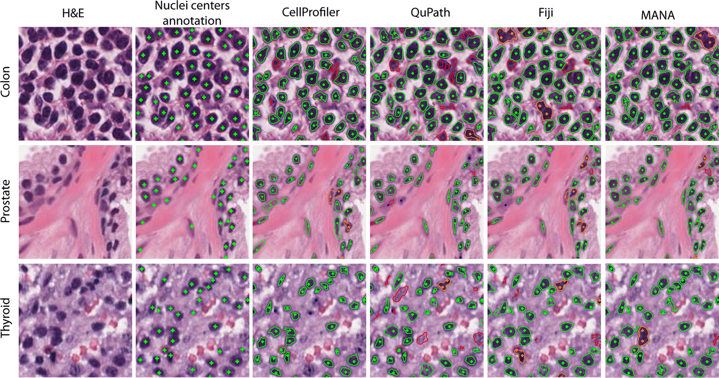

Lighting, and ground-truth depth maps can be found at the Middleburyĭ. The original resolution images, images with different exposure and NaNs denote pixels in the left image that do not have ground-truth. Ground-truth disparity map, where each value describes the offset inĬolumn direction between corresponding pixels in the left and the right In the following and the included ground-truth disparity map are valid forįocal length : 994.978 px Principal point x : 311.193 px Principal point y : 254.877 px Principal point dx : 31.086 px Baseline : 193.001 mm Returns img_left (500, 741, 3) uint8 ndarray

The images are down-sampled by a factor of 4 using The images included here are down-sampled versions of the default exposure A detailedĭescription of the acquisition process can be found in. Greg Krathwohl, and Daniel Scharstein at Middlebury College. Theĭataset was created by Nera Nesic, Porter Westling, Xi Wang, York Kitajima, The two images are part of the Middlebury 2014 stereo benchmark. The ground-truth pixel offset inĬolumn direction is specified by the included disparity map. The background shares enough grey levels with the coins that a simple segmentation is not sufficient. segmentation tests, where individual objects need to be identified against a background. That both images are warped such that they have the same orientation but a This image shows several coins outlined against a gray background. Its corresponding pixel on the same scanline in the right image. The two images are rectified such that every pixel in the left image has Gray-level “text” image used for corner detection.Ĭase B1 image pair from the first PIV challenge. Rectified stereo image pair with ground-truth disparities. Microscopy image of dermis and epidermis (skin layers). Launch photo of DSCOVR on Falcon 9 by SpaceX.

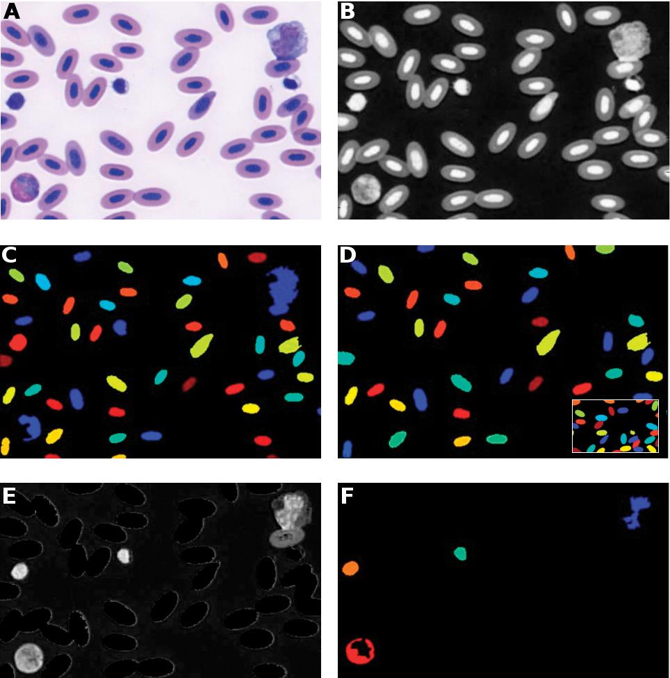
Microscopy image sequence with fluorescence tagging of proteins re-localizing from the cytoplasmic area to the nuclear envelope. Image sequence of synchrotron x-radiographs showing the rapid solidification of a nickel alloy sample. Return the path to the XML file containing the weak classifier cascade. Immunohistochemical (IHC) staining with hematoxylin counterstaining. Measure the granularity, texture, intensity, size and shape. Subset of data from the University of North Carolina Volume Rendering Test Data Set.ģD fluorescence microscopy image of cells.ĭownload all datasets for use with scikit-image offline. Remove the foreground from the original image. Generate synthetic binary image with several rounded blob-like objects. Color image of the astronaut Eileen Collins.


 0 kommentar(er)
